

Deep Phenotyping
Group Leader:
Prof John Le Quesne
The deep phenotyping team are part of the Le Quesne laboratory, and we provide access to advanced spatial biology technologies to our collaborators. The best results come from working very closely with our collaborators in experimental design, and then through the optimisation process, to generate data with the most power to answer the underlying scientific questions. Once data are generated, typically in the form of extremely data-rich images, there are a number of options: we are powerfully equipped to collaborate in digital image analysis and quantification, or to train collaborators to use digital image analysis platforms, but equally often our collaborators will make use of raw data for their own pipelines. Our greatest strengths lie in the design and application of highly multiplexed assays for the detection of RNA and protein in intact tissues. Many of our assays directly address key questions in the immune microenvironment, cellular plasticity, and cell-cell interactions, but we are constantly developing new methods and assays to answer bespoke scientific questions. We enjoy a challenge!
Key technologies
Multiplex immunofluorescence
Using our Ventana Discovery Ultra autostainers in combination with the Akoya PhenoImager HT, we routinely detect up to 6 genes of interest on the same tissue section, allowing us to answer complex questions about tumour biology in a high-throughput manner. We are leaders in this technology, and have collaborations with leading industry providers as well as numerous academic partners. We believe multiplex assays on this scale are likely to form the basis of the next generation of clinical biomarkers, and are working towards translating our assays into a clinical setting.
Over 2025, we have worked with 14 research groups with this technology, and we have several manuscripts in preparation. We have developed panels for human prostate cancer (Leung, Pearson), human pancreatic ductal adenocarcinoma (Campbell), human colorectal cancer (Roxburgh), mouse models of hepatocellular carcinoma (Bird), cholangiocarcinoma (Braconi), and both human and mouse models of malignant mesothelioma (Chalmers, Murphy).
High-plex immunofluorescence
Using our Akoya PhenoCycler Fusion, we can detect up to 100 proteins of interest within intact tissue context. Our expertise with this platform has led to several large in-depth phenotyping collaborations, including our involvement in the SAMBAI Cancer Grand Challenge. This project is a multi-centre international effort to tackle cancer inequities in minority and socially deprived patient populations. In addition, we have collaborations with the Pearson lab at Cardiff University, focusing on fibroblast subtypes in human prostate cancer, and locally with the Chang, Inman and Le Quesne labs focusing on fibroblast subtyping and immune phenotypes in human malignancies, and the Morton lab in mouse models of pancreatic cancer.
We have a methods paper detailing our in-depth validation protocol in process, and our custom PDAC panel has been presented at several meetings. This panel of 43 markers simultaneously detects expression of numerous markers of epithelial and fibroblast plasticity, new therapeutic targets, and highly detailed immunophenotypes. A murine version of this panel is in preparation.
Xenium
Our 10x Genomics Xenium platform enables detection of up to 5,100 genes of interest in a single tissue section. The custom gene expression chemistry has been used by our collaborators to develop panels detecting multiple species simultaneously in parasite-infected animal tissues, and also to detect single nucleotide variants in mRNA. Furthermore, the Xenium can be combined with PhenoCycler Fusion, enabling extremely high-plex multi-omic detection of RNA and protein within the same tissue section. Of note, the Xenium is part of our SAMBAI Cancer Grand Challenge work package.
The Xenium is a key technology for the CRC STARS project, and data from our first mouse custom gene expression panel has been published in Nature by the Sansom lab.
Visiopharm
Visiopharm is our image analysis platform of choice. We have developed bespoke deep learning pipelines for several diseased tissues and tumour types, supporting high-throughput analysis of the images generated by our spatial platforms. We offer training for collaborators wanting to generate their own data, or we design custom workflows and run them as part of our deep phenotyping service.
This year we have published a manuscript in PLOS ONE describing a novel deep learning classifier we have developed using the deep learning tools within Visiopharm, which accurately predicts cellular phenotypes without the need for accurate cell boundary classification.
Lunaphore COMET
The Lunaphore COMET enables the detection of up to 40 proteins, or 12 RNAScope targets and 24 proteins, on the same tissue section. We have established both human and murine immuno-oncology panels for our collaborators to build on. We have also developed multiomic panels for the Sansom and Dasgupta labs.
Visium
This year, we have added the Visium to our facility, offering whole transcriptome analysis using the CytAssist. This can be used as a standalone technology, or in combination with any of the platforms mentioned above.
Multiplex immunochromogenic assays
As a team we are engaged in the translation of our multiplex and high-plex research assays to the clinic, where they can help answer complex diagnostic questions, and ultimately be used as clinical biomarkers in the future.
We believe lower-plex chromogenic multiplex assays using 3 or 4 dyes visible by eye are the way to achieve this, and we have several collaborations in this area.

Recent Publications
click here to read

Lab Members
click here to read
Recent Publications
2026
Pardo, L., Moore, M., Deshmukh, R., Powley, I., Waldron, J.A., Kruspig, B., McGarry, L., Dolma, L., Campos, A.V., Wood, C., Leslie, H., Hughes, M., Jeldes, E., Munro, J., Mitchell, L., Officer-Jones, L., Baird, R., Coquelet, H., Jamieson, N.B., Sumpton, D., Strathdee, D., le Quesne, J., Bushell, M., Murphy, D.J., Norman, J.C., 2026. Increased mRNA translation delays tumour initiation and exposes a therapeutic vulnerability in lung cancer. Mol Cancer.
Williams, H.L., Poulain, N., Powley, I., Martinelli, S., Bielik, R., Leslie, H., Nixon, C., Wilson, C.R., Sereno, M., He, Z., Officer-Jones, L., Ballantyne, F., Pennie, R., Wood, C.S., Lewis, D.Y., Jamieson, N.B., Le Quesne, J., 2026. Spatial Molecular Plasticity Underpins Lethal Morphologies in Lung Adenocarcinoma. Mod Pathol 100960.
2025
Baird, R.L., Mason, D., Rakovic, K., Ballantyne, F., Powley, I.R., Georgakopoulou, A., Hillary, L., Bird, T.G., Officer-Jones, L., Le Quesne, J., 2025. PixlMap: A generalisable pixel classifier for cellular phenotyping in multiplex immunofluorescence images. PLoS One 20(12), e0317865.
Kanellos, G., Giacomelli, C., Raven, A., Vlahov, N., Jin, H., Herviou, P., Malla, S.B., Nasreddin, N., Centeno, P.P., Alexandrou, C., Gilroy, K., Baird, R.L., Pennel, K., Munro, J., Waldron, J.A., Hall, H., Officer-Jones, L., Bryson, S., Strathdee, D., Lilla, S., Zanivan, S., Morrison, V., Nixon, C., Ridgway, R.A., Miller, C., Knight, J.R.P., Campbell, A.D., Dunne, P.D., Le Quesne, J., Edwards, J., Park, P.J., Bushell, M., Sansom, O.J., 2025. Nucleophosmin supports WNT-driven hyperproliferation and tumor initiation. Nat Genet 58, 100–115.
Müller, M., May, S., Hall, H., Kendall, T.J., McGarry, L., Blukacz, L., Nuciforo, S., Georgakopoulou, A., Jamieson, T., Phinichkusolchit, N., Dhayade, S., Suzuki, T., Huguet-Pradell, J., Powley, I.R., Officer-Jones, L., Pennie, R.L., Esteban-Fabró, R., Gris-Oliver, A., Pinyol, R., Skalka, G.L., Leslie, J., Hoare, M., Sprangers, J., Malviya, G., Mackintosh, A., Johnson, E., McCain, M., Halpin, J., Kiourtis, C., Nixon, C., Clark, G., Clark, W., Shaw, R., Hedley, A., Drake, T.M., Tan, E.H., Neilson, M., Murphy, D.J., Lewis, D.Y., Reeves, H.L., Le Quesne, J., Mann, D.A., Carlin, L.M., Blyth, K., Llovet, J.M., Heim, M.H., Sansom, O.J., Miller, C.J., Bird, T.G., 2025. Human-correlated genetic models identify precision therapy for liver cancer. Nature.
Seyedshahi, F., Rakovic, K., Poulain, N., Claudio Quiros, A., Powley, I.R., Richards, C., Uraiby, H., Klebe, S., Moore, D.A., Nakas, A., Wilson, C.R., Sereno, M., Officer-Jones, L., Ficken, C., Teodosio, A., Ballantyne, F., Murphy, D., Yuan, K., Le Quesne, J., 2025. A histomorphological atlas of resected mesothelioma discovered by self-supervised learning from 3446 whole-slide images. Nat Commun 16(1), 8891.
White, M., Mills, M.L., Millett, L.M., Gilroy, K., Hong, Y., Zeiger, L.B., Simpson, R.J., Corry, S.M., Ligeza, A., Lannagan, T.R.M., Susanti, S., Ridgway, R.A., Yazgili, A.S., Grzesiak, L., Amirkhah, R., Ford, C.A., Vlahov, N., Tovell, H., Officer-Jones, L., Ficken, C., Pennie, R., Najumudeen, A.K., Raven, A., Nasreddin, N., Chauhan, E., Papanastasiou, A.S., Nixon, C., Morrison, V., Jackstadt, R., Graham, J.S., Miller, C.J., Ross, S.J., Barry, S.T., Pavet, V., Wilson, R.H., Le Quesne, J., Dunne, P.D., Tejpar, S., Leedham, S., Campbell, A.D., Sansom, O.J., 2025. MAPK-driven epithelial cell plasticity drives colorectal cancer therapeutic resistance. Nature.
2024
Williams, H.L., Poulain, N., Powley, I., Martinelli, S., Bielik, R., Leslie, H., Nixon, C., Wilson, C.R., Sereno, M., He, Z., Officer-Jones, L., Ballantyne, F., Pennie, R., Wood, C.S., Lewis, D.Y., Jamieson, N.B., Le Quesne, J., 2024. Spatial resolution of transcriptomic plasticity states underpinning lethal morphologies in lung adenocarcinoma. bioRxiv.
Xavier, V., Martinelli, S., Corbyn, R., Pennie, R., Rakovic, K., Powley, I.R., Officer-Jones, L., Ruscica, V., Galloway, A., Carlin, L.M., Cowling, V.H., Le Quesne, J., Martinou, J.C., MacVicar, T., 2024. Mitochondrial double-stranded RNA homeostasis depends on cell-cycle progression. Life Sci Alliance 7.

Senior Scientific Officer
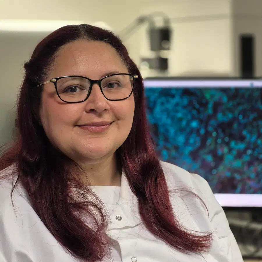
Catherine Ficken
I’ve worked in Histology for over 26 years and I am currently a Senior Scientific Officer in the Deep Phenotyping Advanced Technology Facility. My role focuses on developing high-plex multiplex assays using an array of spatial technologies and I am the technical lead for the 10X Genomics Xenium platform. Outside of the lab, you’ll usually find me at a gig singing my heart out, curled up with a delightfully trashy romance novel, watching a good film, or catching up with friends over a cocktail or two putting the world to rights.
Senior Scientific Officer
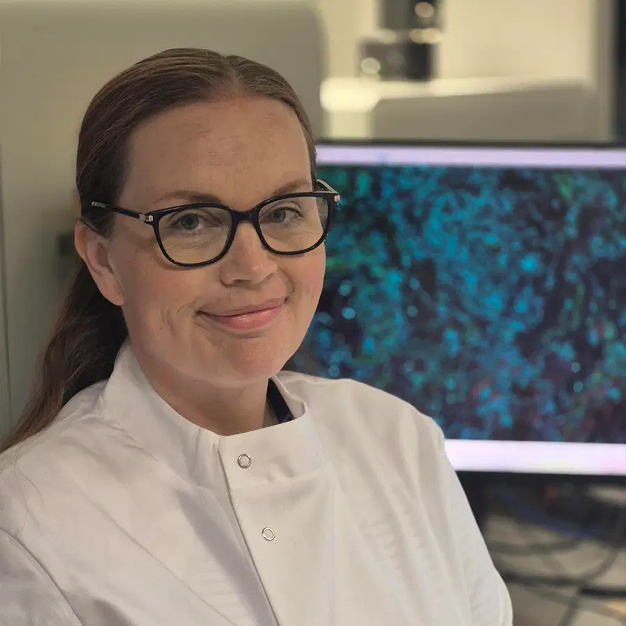
Denise McPhee
I am a Senior Scientific Officer in the Deep Phenotyping Advanced Technology Facility. I have almost 20 years' experience in diagnostic and research histology, with a particular interest in immunohistochemistry and quality assurance processes. Within this team, I work with the Visium platform, develop multiplex panels and I am also the technical lead for the Ventana platform. Outside of work I enjoy reading, the outdoors, and spending time with my husband, children and animals.
Senior Scientific Officer

Fiona Ballantyne
I’m a Senior Scientific Officer with a BSc in Anatomy from University of Glasgow. I am currently the technical lead for the Lunaphore COMET and the Akoya PhenoCycler where I develop high-plex proteomic and multiomic panels for researchers.
Scientific Officer
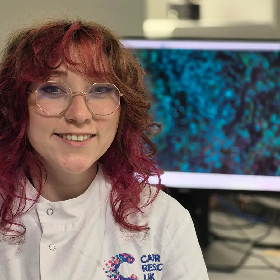
Kara Luckett
Before I became a Scientific Officer in the Deep Phenotyping Advanced Technology Facility, I received my BSc in Forensic Biology at West Virginia University and MSc (MedSci) in Medical Genetics at the University of Glasgow. I spend my time optimising high-plex spatial biology panels. When not in the lab, you can find me hiking in the woods, burrowed into a good book, or front row at a concert.
Histopathology Technology Manager
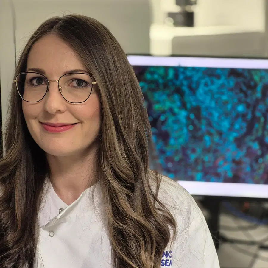
Leah Officer-Jones
I manage the Deep Phenotyping Advanced Technology Facility. I started out as a Cellular Pathology Scientist, and I have an MSc in Molecular Pathology of Cancer from Queen’s University Belfast. I have spent the last 10 years developing multiplex and spatial biology methods. Day to day I manage a team of expert translational histologists, and a facility housing several multiplex and spatial biology technologies. When I’m not at work you’ll find me in my garden, or exploring the highlands and islands with my paddleboard.
Scientific Officer
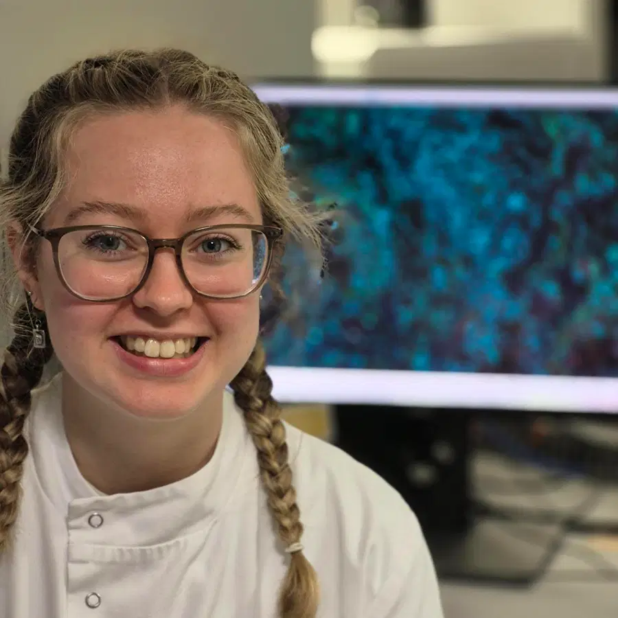
Lucy Hillary
I’m Lucy, a Scientific Officer within the Deep Phenotyping Advanced Technology Facility. I have recently graduated from the University of Glasgow with an MSci in Molecular and Cellular Biology, where I completed a placement year within the Le Quesne Laboratory. During my placement, I conducted a spatial biology project, focusing on fibroblast subtyping in thoracic malignancies. Outside of the lab, I enjoy playing squash, indoor bouldering, and reading.
Senior Scientific Officer
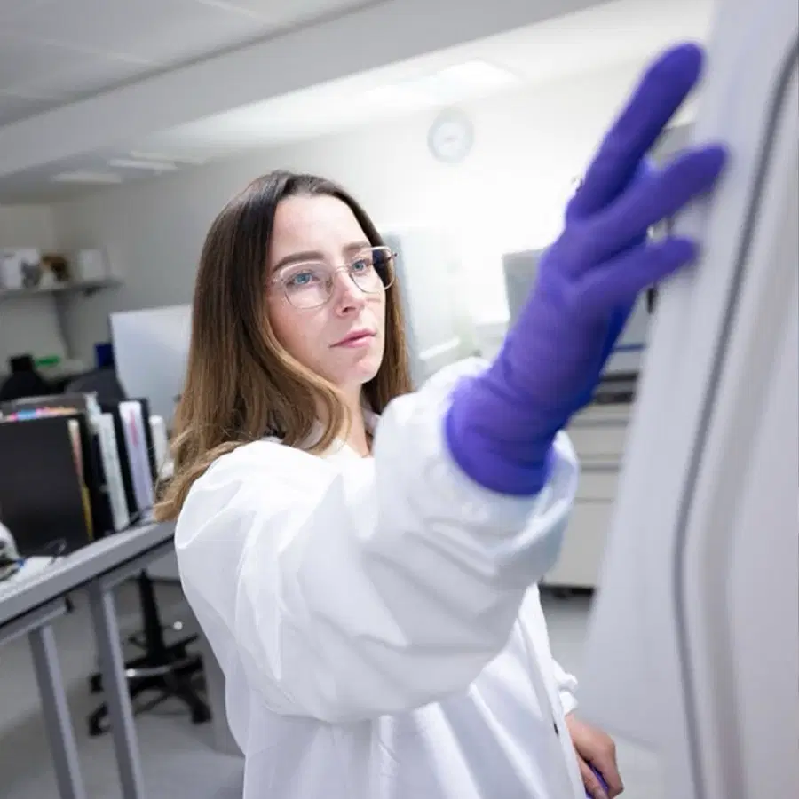
Rachel Baird
I am a Senior Scientific Officer within the Deep Phenotyping Advanced Technology Facility, where I work as a translational histologist. My expertise lies in developing advanced multiplex assays and creating innovative image analysis workflows for multiplex and high-plex imaging. I hold a BSc in Pharmacology, and an MSc in Cancer therapies with the University of Strathclyde. Outside of the lab, you’ll find me going for a coffee and a cake with my husband, daughter, and dog, and (maybe) going to the gym during the day, with a glass of wine by night.