

Beatson Advanced Imaging Resource
Group Leader:
Prof Leo Carlin
Light microscopy allows us to gather information about important regulatory mechanisms in tumours and the microenvironment. Using these techniques, we can simultaneously analyse large numbers of important molecules and cells with subcellular sensitivity and resolution in living samples whilst maintaining the context of the microenvironment, be that model substrate or living organism.
The Beatson Advanced Imaging Resource (BAIR) team works closely with the Institute’s researchers to uncover and interrogate important molecular pathways in cancer. The BAIR is thus involved at some stage in nearly every study from researchers at the Institute that contains a light micrograph. We are keen and able to assist from experimental design right through to the finished figures. We train scientists in all stages of modern cytometric and microscopical research, from advice and help with sample preparation, basic and advanced microscope and cytometer operation, and data acquisition through to quantitative image analysis and interpretation. At the start of a new project or application, we are enthusiastic to help researchers identify how our methods can be used to develop and test their hypotheses and help them to design experiments that make the most of our advanced instrumentation. We also identify and acquire new technology and methodology that allow our researchers to take the most elegant approaches.
Imaging across different spatial and biological complexity scales
We have the expertise and instruments to:
- Perform automated liquid / multi-well plate handling and very high-throughput imaging experiments to analyse cell behaviour over thousands of experimental conditions via high-content imaging
- Image, spatially separate, and quantify up to eight markers simultaneously in thick tissue (3 and 4D) by combining fluorescently labelled antibodies and probes with label-free approaches (e.g. second harmonic generation to look at fibrillar collagen) using tissue clearing, multiphoton excitation and spectral imaging
- Image cell behaviour over several days in tissue culture incubators
- Address the physicochemical environment, molecular activity, and signal transduction of pathways below the diffraction limit at different spatiotemporal scales using FLIM, FRET and super-resolution imaging
- Monitor cell function in intact living organisms via advanced intravital microscopy
In this way, we underpin cancer research at the Institute, UoG School of Cancer Sciences and beyond by allowing our researchers to work up and down the biological complexity scale, taking the best and most important aspects of different models and patient samples and combining them into a larger more complete picture.

Recent Publications
click here to read

Lab Members
click here to read
Recent Publications
2026
Riggio, A.I., Sweeney, K., Shaw, R., Lawlor, A., Khan, A., Ferrari, N., Anand, J., Galbraith, L.C., Gilroy, K., Bull, C., Young, A.L., Athineos, D., Hall, H., Ghaffar, F., Hughes, M., Mitchell, C.A., Mitchell, L., Nixon, C., Adams, P.D., Roberts, E.W., Miller, C.J., Dunne, P.D., Campbell, K.J., Cameron, E.R., Blyth, K., 2026. Runx1 and Runx2 act in concert to suppress Wnt/β-catenin-driven mammary tumourigenesis. Br J Cancer 1–14.
Raffo-Iraolagoitia, X.L., McFarlane, A.J., Laing, S., Corbyn, R., Arnott, L.W.G., Fercoq, F., McGarry, L., Secklehner, J., De Donatis, M., Mackey, J.B.G., Kruspig, B., Wiesheu, R., Hsieh, Y.-C., Shaw, R., Rakovic, K., Le Quesne, J., Clark, G., Nixon, C., Miller, C., Kirschner, K., Bain, C.C., Murphy, D.J., Coffelt, S.B., Carlin, L.M., 2026. Vɣ1 ɣδ T cells steer airway macrophages toward a profibrotic response in an autochthonous lung cancer mouse model. Science Advances 12, eadu8802.
Pardo, L., Moore, M., Deshmukh, R., Powley, I., Waldron, J.A., Kruspig, B., McGarry, L., Dolma, L., Campos, A.V., Wood, C., Leslie, H., Hughes, M., Jeldes, E., Munro, J., Mitchell, L., Officer-Jones, L., Baird, R., Coquelet, H., Jamieson, N.B., Sumpton, D., Strathdee, D., le Quesne, J., Bushell, M., Murphy, D.J., Norman, J.C., 2026. Increased mRNA translation delays tumour initiation and exposes a therapeutic vulnerability in lung cancer. Mol Cancer.
Fercoq, F., Cairns, G.S., Bridgeman, V.L., De Donatis, M., Mackey, J.B.G., Floerchinger, A., McFarlane, A.J., Secklehner, J., Raffo-Iraolagoitia, X.L., Harrington, M., McGarry, L., Brownlie, D., Whyte, D., Arnott, L.W.G., Nixon, C., Wiesheu, R., Kilbey, A., Brown, L., Al-Khalidi, S., Norman, J.C., Roberts, E.W., Malanchi, I., Blyth, K., Coffelt, S.B., Carlin, L.M., 2026. Integrin-dependent neutrophil slowing reduces lung perfusion and supports metastasis in a model of breast cancer. Cell Reports 45, 117101.
Dunlop, J.I., Thomason, P.A., Carlin, L.M., Davis, B.G., Carter, S.D., 2026. Live-cell 3D-SIM of Rift Valley fever virus NSs filaments reveals a polygon web architecture. Proceedings of the National Academy of Sciences 123, e2534404123.
2025
Müller, M., May, S., Hall, H., Kendall, T.J., McGarry, L., Blukacz, L., Nuciforo, S., Georgakopoulou, A., Jamieson, T., Phinichkusolchit, N., Dhayade, S., Suzuki, T., Huguet-Pradell, J., Powley, I.R., Officer-Jones, L., Pennie, R.L., Esteban-Fabró, R., Gris-Oliver, A., Pinyol, R., Skalka, G.L., Leslie, J., Hoare, M., Sprangers, J., Malviya, G., Mackintosh, A., Johnson, E., McCain, M., Halpin, J., Kiourtis, C., Nixon, C., Clark, G., Clark, W., Shaw, R., Hedley, A., Drake, T.M., Tan, E.H., Neilson, M., Murphy, D.J., Lewis, D.Y., Reeves, H.L., Le Quesne, J., Mann, D.A., Carlin, L.M., Blyth, K., Llovet, J.M., Heim, M.H., Sansom, O.J., Miller, C.J., Bird, T.G., 2025. Human-correlated genetic models identify precision therapy for liver cancer. Nature 639, 754–764.
Gounis, M., Campos, A.V., Shokry, E., Mitchell, L., Deshmukh, R., Dornier, E., Rooney, N., Dhayade, S., Pardo, L., Moore, M., Novo, D., Mowat, J., Jamieson, C., Kay, E., Zanivan, S., Paul, N.R., Mitchell, C., Nixon, C., Macpherson, I., Tardito, S., Sumpton, D., Blyth, K., Norman, J.C., Clarke, C.J., 2025. Metabolic adaptations of micrometastases alter EV production to generate invasive microenvironments. J Cell Biol 224, e202405061.
2024
Xavier, V., Martinelli, S., Corbyn, R., Pennie, R., Rakovic, K., Powley, I.R., Officer-Jones, L., Ruscica, V., Galloway, A., Carlin, L.M., Cowling, V.H., Quesne, J.L., Martinou, J.-C., MacVicar, T., 2024. Mitochondrial double-stranded RNA homeostasis depends on cell-cycle progression. Life Science Alliance 7.
Mandrou, E., Thomason, P.A., Paschke, P.I., Paul, N.R., Tweedy, L., Insall, R.H., 2024. A Reliable System for Quantitative G-Protein Activation Imaging in Cancer Cells. Cells 13, 1114.
Burgess, S.G., Paul, N.R., Richards, M.W., Ault, J.R., Askenatzis, L., Claydon, S.G., Corbyn, R., Machesky, L.M., Bayliss, R., 2024. A nanobody inhibitor of Fascin-1 actin-bundling activity and filopodia formation. Open Biology 14, 230376.
Group Member
Peter Thomason
My training is in biochemistry and cell biology. During several post-docs and fellowships I investigated intra- and inter-cellular signaling, mostly in the context of cell migration and development. Latterly I focused on the role of the cytoskeleton in cell movement and trafficking of vesicles. Through these projects I became deeply involved in advanced microscopy techniques, learning them from the experimenter’s perspective. My expertise encompasses live confocal, spectral, and super-resolution (Airyscan and SIM) imaging.
Principal Scientific Officer
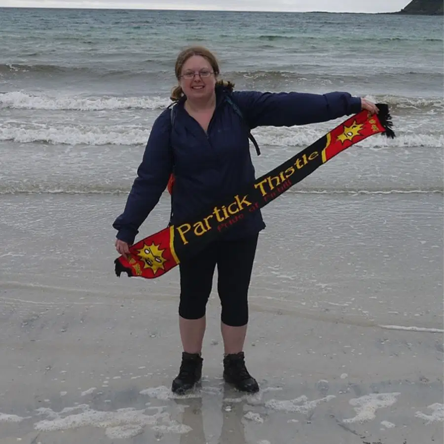
Nikki Paul
work as a principal scientific officer in BAIR, and I have worked in the CRUK Scotland Institute since 2015. I have a background in live and fluorescence microscopy of cell migration and metastasis, and have worked on cancer models both at CRUK-SI and during my PhD at the University of Manchester. My background is in Biochemistry with a focus on cell biology.
In BAIR I specialise in sample optimisation, live-imaging and super-resolution microscopy. As co-chair of the Scottish Microscopy Society (SMS) I aim to improve networking opportunities for microscopy across the country. I am also involved in public engagement and science communication. In my spare time I like exploring Scotland, birds and wildlife, and sports, especially football.
Senior Scientific Officer
Ryan Corbyn
I am a Senior Scientific Officer within the Beatson Advanced Imaging Resource at the CRUK Scotland Institute. My primary role in the team is as an image analyst. I help create bespoke workflows for the research groups within the institute to gain as much insight as possible from their imaging experiments. Before joining the BAIR team, I completed a PhD at the University of Strathclyde, where I developed a confocal microscope to detect biologically generated magnetic fields using quantum sensing protocols.
Outside of the institute, my hobbies include bouldering/climbing, hiking, and learning to play the guitar.
Principal Scientific Officer
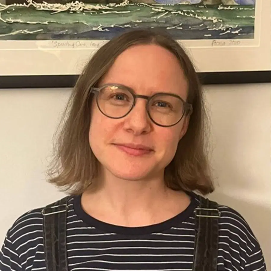
Claire Mitchell
I work as a principal scientific officer in BAIR. I have worked in light microscopy facilities since 2017 and am experienced in most light microscopy techniques. My background is as an optical engineer, completing a Physics PhD at the University of St Andrews. My role in BAIR is working with our advanced systems, including multiphoton and light sheet. I am also passionate about quality control, reproducibility and education in microscopy. In my spare time I like to be outdoors, going camping, hiking, paddleboarding and foraging.
Senior Scientific Officer
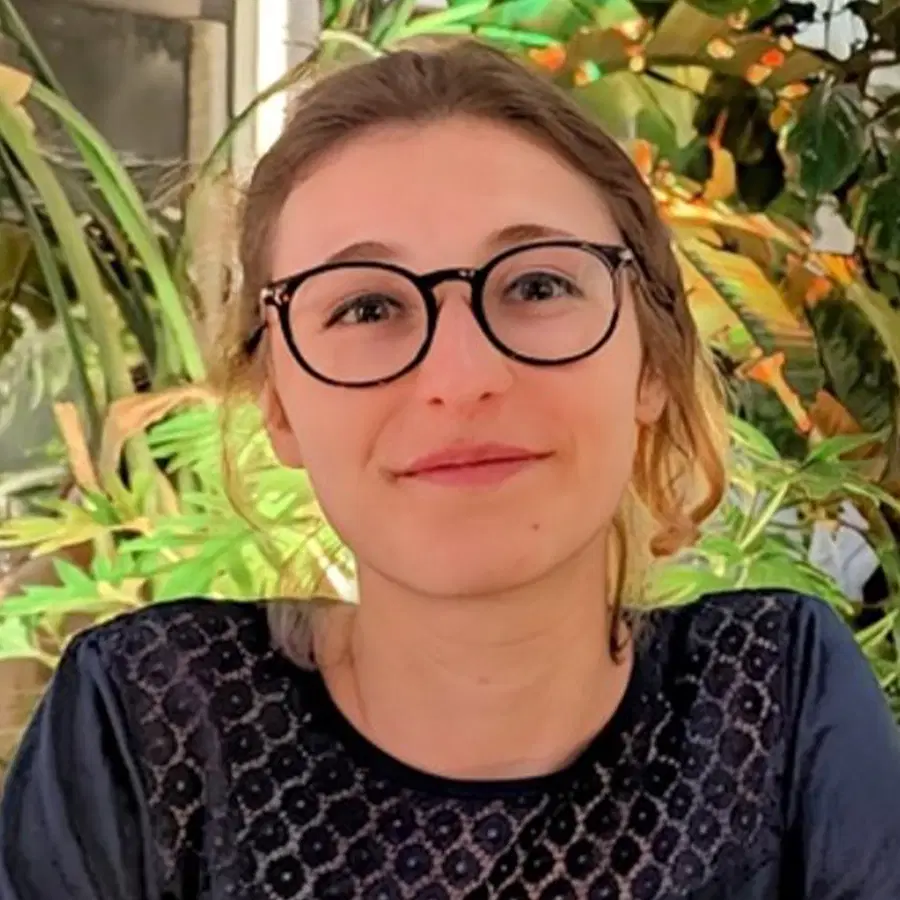
Beatrice Bottura
I was born in Italy but spent most of my school years in France, then moved to England for a MPhys degree in Physics and finally to Glasgow for a PhD in Strathclyde's Mesolab. There I discovered my passion for both microscopy and biology (especially wet lab work). As a Senior Scientific Officer in BAIR, I support the High-Content / High-Throughput Screening facility, helping researchers identify genetic and chemical vulnerabilities in different types of cancer. Outside of work, I love exploring new places (both in Scotland and around the world), cooking and catching up with my friends during our weekly D&D campaign.
Senior Scientific Officer
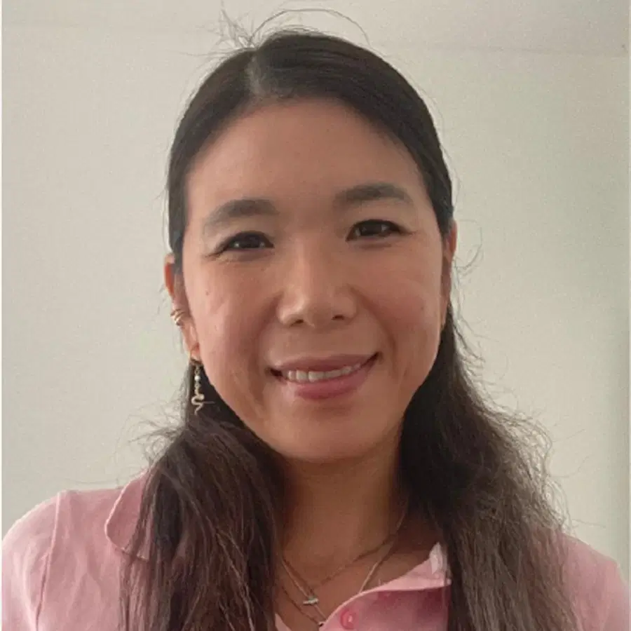
Jinyi Su
I am a Senior Scientific Officer at BAIR since March 2026. Before joining CRUK-SI, I completed my PhD in Infection Biology at Utrecht University. My main areas of expertise are molecular cell biology, gut biology, and host–pathogen interactions. I am passionate about New Approach Methodologies (NAMs), data analysis, and data management. Together with Beatrice, we run and support high-throughput and high-content screening for the team. We assist researchers and scientists throughout the entire process, from hypothesis testing and experimental design to execution and data analysis. Outside of the lab, I enjoy reading a good book, watching films, exploring the city (especially independent cafés and bakeries), and hiking with dogs.